View Silver Stain Collagen US
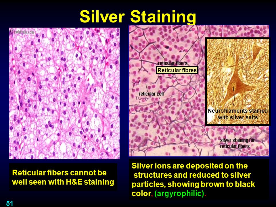
View Silver Stain Collagen US. Silver staining aids the visualization of targets of interest, namely intracellular and extracellular cellular components such as dna and proteins, such as type iii collagen and reticulin fibres by the deposition. (* collagen may not stain uniformly as the dye take up depends on the physical state of the fibers silver impregnation carried out in ammonical silver nitrate solution ↓ reduction with formaldehyde.

The procedures of silver staining are protein fixation, silver impregnation and image development.
Trichrome and van gieson stains can show all types of collagen. Collagen may be stained with light green instead of aniline blue. Cytoplasm (including erythrocytes) and nucleoli. Bio134 stain collagen, elastic and reticular fibers.
Komentar
Posting Komentar